Bronchopulmonary Sequestration: What It Is And What Does It Consist Of
Bronchopulmonary sequestration refers to a structural alteration of the lungs in which a part of the lung structure or parenchyma is isolated from the rest of the respiratory system, so it does not function properly. It is a rare congenital malformation.
It accounts for 2% of lung resections (surgeries in which part of the lung is removed). There are two types of bronchopulmonary sequestration: intralobar and extralobar.
Let’s see more below.
Physiology of the respiratory system
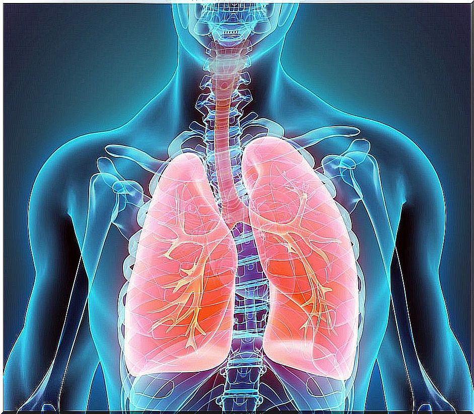
The main function of the respiratory system is to allow oxygen to enter the body. It is a vital requirement for proper cell function. It is achieved thanks to the gas exchange that takes place in the lungs, in which oxygen enters and carbon dioxide is expelled.
The organs that make up the respiratory system are the nasal passages, pharynx, larynx, trachea, bronchi, bronchioles, and lungs.
For gas exchange to take place, the blood must reach the lungs filled with carbon dioxide, which is a waste product of cellular activity. The artery that carries blood to the lungs is the pulmonary artery, which exits the heart.
It is in structures called alveoli that the exchange of oxygen for carbon dioxide takes place. The pulmonary veins then carry the oxygenated blood back to the heart, from where it is distributed to the rest of the body to deliver oxygen to the tissues.
Pathogenesis of bronchopulmonary sequestration
There are multiple theories about the etiopathogenesis of bronchopulmonary sequestration. It is believed that during embryonic lung development an accessory pulmonary diverticulum forms that grows independently of the rest of the tracheobronchial tree.
As we have said, there are two varieties of bronchopulmonary sequestration. To understand the difference we must remember that the lungs are asymmetric and are divided into lobes:
- Right lung: it is larger and consists of three lobes, upper, middle and lower.
- Left lung: only two lobes, upper and lower. It is smaller in size by sharing space in the thoracic cavity with the heart.
In turn, the lobes are divided into segments. It is also interesting to remember that they are surrounded by a serous layer called the pleura . So, depending on the moment of development in which this diverticulum emerges, two things can happen:
- If the bud forms before the formation of the pleura, when it develops, it will also envelop the bud, giving an intralobar sequestration.
- If it is formed after the development of the pleura, the sprout grows independently. Thus it is enveloped in its own serous layer. We speak then of extralobar kidnapping.
Intralobar bronchopulmonary sequestration
It’s the most frequent form. It makes up about 75% of lung sequestration. It is usually located in the lower lobes. It shares the pleura with the rest of the lung, a fact that differentiates it from extralobar pulmonary sequestration.
It receives blood from systemic arteries, mainly from branches of the aorta, but venous drainage is carried out through the pulmonary vein.
Extralobar bronchopulmonary sequestration

Extralobar pulmonary sequestration is less frequent, accounting for 25% of cases. In this case, the lung fragment has its own pleura and may be separated from the rest of the lung. The most common locations are the mediastinum, the pericardium, or below the diaphragm.
Irrigation is usually at the expense of some aortic branch. In this case, venous drainage is not done through the pulmonary vein but is also systemic. It can lead to the subclavian, azygous or portal vein.
Bronchopulmonary Sequestration Clinic
The clinical manifestations of this malformation will depend on the type of sequestration and whether it associates other alterations. In fact, in the case of intralobar sequestration there are usually no symptoms, it is diagnosed as an incidental finding when a chest X-ray is performed for another reason. When symptoms appear, they are not specific, the most common are:
- Fever.
- Chest pain
- Productive cough with expulsion of mucus.
- Feeling of shortness of breath or shortness of breath.
Although the “sequestered” segment is not functionally connected to the airway, inhaled microorganisms can reach it through communications established in the bronchioles, which is why respiratory infections are common.
Diagnosis of bronchopulmonary sequestration
While the intralobar type is usually asymptomatic and is diagnosed in advanced ages, the extralobar type is usually diagnosed in the neonatal period. Among other things, this is because it is associated with other congenital malformations.
Diagnosis can be in utero or at any time in development. It is based on imaging techniques such as chest x-ray or ultrasound. The image will show a condensation similar to pneumonia.
Confirmation of the diagnosis
To confirm the diagnosis, CT angiography is used, which is the gold standard of this test since it demonstrates the independent vascularization of the pulmonary segment. This fact allows it to be differentiated from other pulmonary condensations, such as:
- Infections, such as pneumonia and tuberculosis.
- Mediastinal or lung masses.
- Other congenital malformations.
And the treatment?
Treatment consists of surgical removal or resection of the “sequestered” lung territory. Surgery is recommended even in asymptomatic cases since it reduces the risk of infections.